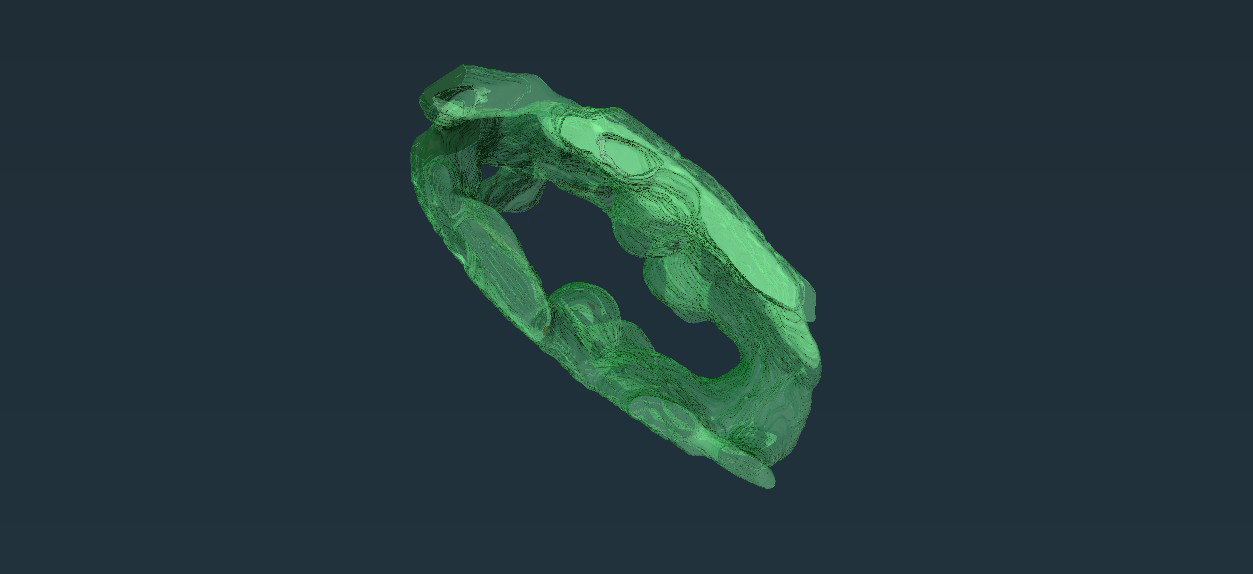
The researchers were able to reconstruct the three-dimensional shape of the single chloroplast from several hundred images. Credit: University of Oldenburg / General and Molecular Microbiology group
A distinctive photosynthetic machinery characterizes a unicellular organism present in algal blooms.
What are the cellular mechanisms within a single-celled marine algae species responsible for triggering toxic algal blooms? A research group under the direction of microbiologist Prof. Dr. Ralf Rabus from the University of Oldenburg, Germany, has conducted first detailed analyses of the unusual cell biology of Prorocentrum cordatum, a globally widespread species of the dinoflagellates group, using both advanced microscopic and proteomics approaches.
As the team reports in the science journal Plant Physiology, the photosynthesis process in these microorganisms is organised in an unusual configuration which may help them to better adapt to the changing light conditions in the oceans. The results of the study could lead to an improved understanding of the incidence of harmful algal blooms, which may be becoming more frequent due to climate change.
Dinoflagellates are important organisms in both marine and freshwater ecosystems. These unicellular organisms make up a substantial proportion of free-living phytoplankton, which forms the basis of the food web in oceans and lakes. Some species, including Prorocentrum cordatum, can proliferate in warm, nutrient-rich waters and form harmful algal blooms.
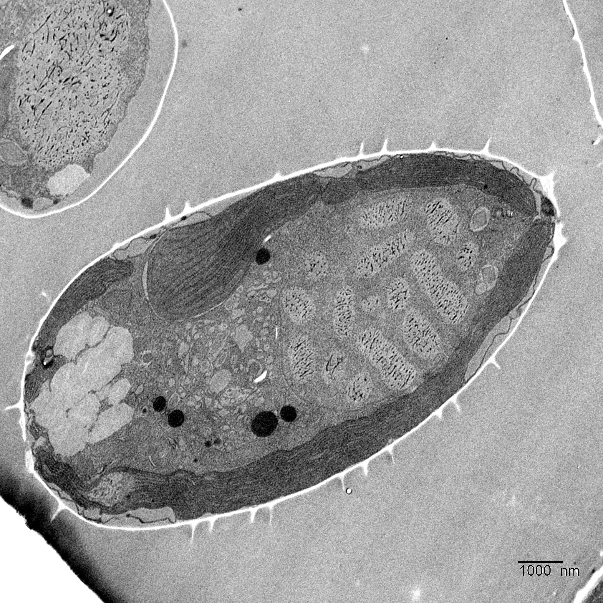
Cross-section of a cell of the microalga Prorocentrum cordatum. The nucleus with the chromosomes is on the right. A single barrel-like chloroplast takes up 40 percent of the cell volume. Credit: University of Oldenburg / General and Molecular Microbiology group
“We studied this organism because despite its environmental relevance its cell biology and metabolic physiology are still poorly understood,” said Rabus. In addition to studying photosynthesis in the microalgae, the researchers also examined the structure of their cell nuclei and their response to heat stress in collaboration with teams from the Universities of Hanover, Braunschweig, and Munich and set out the findings in two other recently published papers.
Advanced Imaging Techniques Reveal Unique Cellular Structures
Using a powerful scanning electron microscope with a focused ion beam at the Ludwig-Maximilians-Universität Munich, the team headed by Rabus and lead author Jana Kalvelage from the Institute of Chemistry and Biology of the Marine Environment (ICBM) was able to reconstruct the three-dimensional architecture of the chloroplasts, where photosynthesis takes place. The scientists were able to generate around 600 image layers of a single algae cell and then combine the sections to create a three-dimensional, high-resolution spatial image of the oval-shaped single-celled organisms, which are generally around 10 to 20 thousandths of a millimeter long. The analysis revealed that Prorocentrum cordatum have only a single barrel-like chloroplast that takes up 40 percent of their cell volume.
Proteomic (protein) analyses then revealed marked differences between the photosynthetic apparatus of the microalgae and that of Arabidopsis thaliana, a well-studied model plant in genetics research. In both species, photosynthesis takes place in complex protein structures embedded in the chloroplast’s extensive membrane system.
However, in Prorocentrum cordatum the team observed that the conversion of solar energy into biochemical energy takes place in a single large structure consisting of numerous proteins, known as a “megacomplex”, whereas in the chloroplasts of the plant species, the different steps of photosynthesis occur in spatially separated structures. The team also reported that P. cordatum uses a large number of different pigment-binding proteins to efficiently capture solar energy. “This diversity is a special adaptation to the changing light conditions to which the organism is exposed in the oceans,” Rabus explained.
Exploring Genetic Complexity and Adaptability
Two other studies published last year highlight the microalgae’s unusual biology: in the first a German-Australian team of which the ICBM researchers were also members found that the organisms have a very large genome with twice as many base pairs as in humans. The team also discovered that the algae change their metabolism and their rate of growth decelerates in response to heat stress. In a second publication, the team led by Rabus and Kalvelage described the cell nucleus in greater detail, reporting that P. cordatum has 62 chromosomes, an unusually high number that fills almost the entire cell nucleus. The function of a large proportion of the nuclear proteins that were identified by the researchers is currently unknown, the team observed.
“We have investigated how this important microalgae functions at the molecular level. These findings form the basis for a better understanding of its role in the environment,” Rabus stressed. Further investigations could provide answers to questions such as how the organism’s metabolism reacts to other stress factors – and why the species is able to adapt to such a wide range of environmental conditions, from those in the tropics to those in temperate climates, he explained.
Reference: “Conspicuous chloroplast with light harvesting-photosystem I/II megacomplex in marine Prorocentrum cordatum” by Jana Kalvelage, Lars Wöhlbrand, Jennifer Senkler, Julian Schumacher, Noah Ditz, Kai Bischof, Michael Winklhofer, Andreas Klingl, Hans-Peter Braun and Ralf Rabus, 08 February 2024, Plant Physiology.
DOI: 10.1093/plphys/kiae052
The study was funded by the German Research Foundation.


Be the first to comment on "Scientists Unravel the Unusual Cell Biology Behind Toxic Algal Blooms"